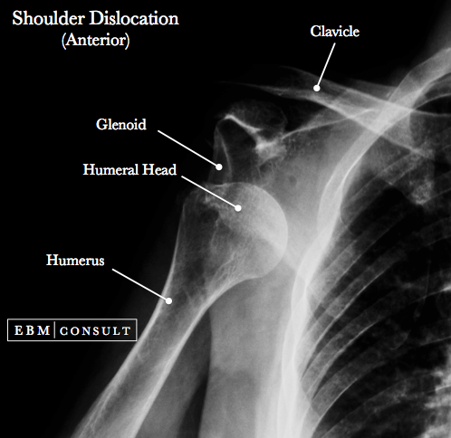
Anterior Shoulder Dislocation
Summary:
- Anterior shoulder dislocations are the most common type of shoulder dislocation and usually occur when the arm is externally rotated and in the abducted position.
- Initial imaging includes plain radiographs of the shoulder: AP and axillary views. Complications include: axillary nerve damage, Bankart lesion, Hill-Sachs lesion, and vascular injuries (though rare).
- Most cases can be reduced using closed (non-surgical techniques) under either local anesthesia or procedural sedation and then placed in a sling with outpatient follow up in 5-7 days with orthopedics.
Anterior Shoulder Dislocation
|
|---|
-
Most common form (~ 98% of shoulder dislocations).
-
Patients will commonly present with the arm externally rotated and in the abducted position.
-
Humeral head can be palpated in anterior shoulder just below the subcoracoid area and clavicle.
-
Classically associated with convulsive seizures and electrocution, though still uncommon.
- Occur with abduction, extension and external rotation where the humeral head is forced out of the glenohumeral joint from detachment of the anterior capsule.
- In elderly patients can occur from fall on outstretched hand.
- Axillary or Scapular "Y" view:
- The humeral head will be lying anterior to, or in front of the glenoid.
- AP of the Shoulder:
- Should see the humeral head in the subcoracoid
position.
- CT Scan or MRI:
- If needing to evaluate for a Hill-Sachs lesion or Bankart lesion.
- Axillary nerve damage (loss of sensation to pinprick in the area of the deltoid).
- Bankart lesion (detachment of the anteroinferior labrum from the glenoid; usually see with MRI).
- Hill-Sachs lesion (a posterolateral humeral head compression fracture when the humeral head is resting against the anteroinferior portion of the glenoid that is associated with an increased risk for future dislocations). Sometimes this can only be seen on a CT scan.
- Vascular injuries: usually rare but consider if pulse deficits present.
-
Check neurovascular status
-
Closed reduction under procedural sedation and/or intra-articular shoulder injection with lidocaine
-
Reduction techniques include: Stimson technique, external rotation method, traction-countertraction method, and scapular rotation
-
After closed reduction, place in sling-and-swath, or shoulder immobilizer
-
Post-reduction films to evaluate for position and presence of fractures (such as a Hill-Sachs lesion)
-
Outpatient follow up with orthopedic surgery within 5-7 days and counsel to minimize activity that would result in abduction and external rotation.
- Radiology: Posterior Shoulder Dislocation
- Kroner K, Lind T, Jensen J.
The epidemiology of shoulder dislocations. Arch Orthop Trauma Surg
1989;108(5):288-90.
- Zhang AL, Montgomery SR, Ngo SS, Hame SL, Wang JC, Gamradt SC. Arthroscopic versus open shoulder stabilization: current practice patterns in the United States. Arthroscopy 2014;30(4):436-43.
Other possible associated injuries include:
To see other related content select from an item below:


