Chest Radiograph - Normal
|
|---|
It is important to know the normal chest radiograph and common landmarks so that you can recognize what is abnormal.

- Right Border: Formed by the right atrium which is in between the SVC and IVC
- Left Border: Formed by the left ventricle & portion of the left auricle
- Anterior Surface or Sternocostal Surface: Mainly the right ventricle (not seen on AP view)
- Inferior Border: Combination of the right & left ventricles
- If they are blunted or lost, you should be concerned for the presence of fluid in the lung or a mass obstructing the view. Additional imaging with a chest CT may sometimes be warranted if the etiology is not clear from the patient's presentation.
- Each dome of the diaphragm is innervated by its own nerve supply from the phrenic nerve. Therefore, damage to the nerves for one side will not affect the other. On chest radiograph you would see the the paralyzed hemidiaphragm as being higher than the other hemidiaphragm during inspiration (creating a paradoxical pattern of movement with respiration).
- You should also not see free air under the hemidiaphragm. If free air is found you will see a black line under the hemidiaphragm which would be concerning for a bowel perforation. This requires emergent evaluation with a CT scan and surgical consult. Do not confuse the normal gastric bubble seem on many chest radiographs with free air.
On an antero-posterior (AP) or postero-anterior (PA) view of the chest, the borders of the heart have common landmarks:
The aortic knob should be visualized in the normal chest radiograph around the level of T3 to T4 or just lateral to the carina. In patients with aortic aneurysm, this can be the area contributing to the "widened mediastinum".
The costocardiac angles (as well as the costophrenic angles) should fairly sharp and well defined if the patient does not have significant effusions or pulmonary edema.
The carina is the point or level at which the trachea divides into the right and left main bronchi. This is usually midline with the spinous process being behind it. The carina is also the location that is used by healthcare providers when assessing the proper position of an endotracheal tube (ET) after intubation. Typically, the tip of the ET tube should be 3-4 centimeters above the carina so that both lungs are properly oxygenated.
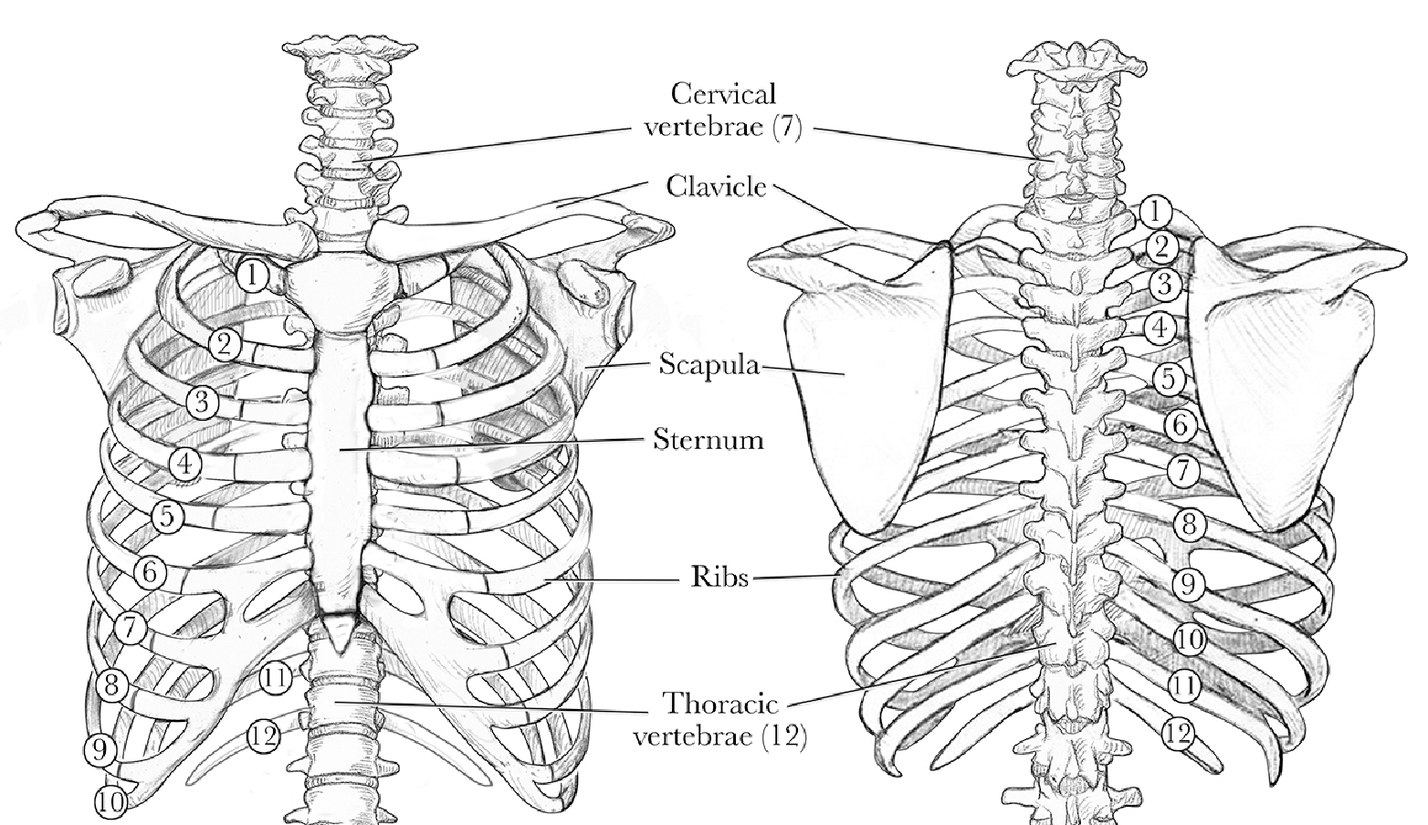
The head of the clavicle is attached to the lateral surface of the sternum. The location of the clavicular heads in relation to the trachea can help determine proper positioning of the patient at the time the chest radiograph was taken. The two clavicular heads should be on either side of the trachea and with the spinous processes being in the middle.
The right hemidiaphragm normally sits slightly higher than the left due to the presence of the liver under the diaphragm which prevents the right hemidiaphragm from going down further with inspiration. Important clinical pearls include:


